Shoulder Ligament Anatomy Diagram : Anatomy Of The Human Shoulder Joint - 3 problems of the shoulder.this acts as the bony framework by which the muscles of the chest, upper back and shoulder connect the.
byAdmin•
0
Shoulder Ligament Anatomy Diagram : Anatomy Of The Human Shoulder Joint - 3 problems of the shoulder.this acts as the bony framework by which the muscles of the chest, upper back and shoulder connect the.. Muscle anatomy atlas 12 photos of the muscle anatomy atlas , human muscles Learn vocabulary, terms, and more with flashcards, games, and other study tools. The shoulder has about eight muscles that attach to the scapula. Shoulder muscles and chest human anatomy diagram pdf arm vertebrate anatomy britannica Located superior to the shoulder joint, the deltoid muscle works with the supraspinatus to abduct the arm at the shoulder.
Anatomy diagrams of shoulder, arm, elbow, forearm, wrist and hand. The long head of biceps (lhb) is a very important tendon that travels through the shoulder joint (glenohumeral joint).the biceps tendon begins at the top of the shoulder socket (the glenoid) and then passes across the front of the shoulder to connect to the biceps muscle. Located superior to the shoulder joint, the deltoid muscle works with the supraspinatus to abduct the arm at the shoulder. 17 photos of the diagram of shoulder muscles and tendons. Diagram of shoulder anatomy showing the acromioclavicular (ac) articulation and glenohumeral (gh) joint.
Dislocated Shoulder Symptoms Causes Treatments from www.clevelandclinic.org The shoulder anatomy includes the anterior deltoid lateral deltoid posterior deltoid as well as the 4 rotator cuff muscles. It contributes to shoulder stability and, when. Bones in shoulder, ligaments of the shoulder joint, parts of the shoulder joint, shoulder anatomy, shoulder joints and muscles, shoulder structure anatomy, shoulder tendon anatomy, shoulder tendons ligaments, human. The anterior shoulder pain usually develops when injury or inflammation occurs in the tendons that are attached to the shoulder joint. You can see the glenoid cavity of the scapula here. The collection of muscles and tendons in the shoulder is known as the rotator cuff. A complete dislocation means the ball comes all the way out of the socket. 3 problems of the shoulder.this acts as the bony framework by which the muscles of the chest, upper back and shoulder connect the.
Beyond this, there is also a shoulder joint arrayed in a ball and socket formation, a rotator cuff, and various muscles like the deltoid muscle and the teres major muscle.
Biceps tendons the biceps muscle has two tendons at the shoulder, called the long head and short head. The shoulder joint is formed where the humerus (upper arm bone) fits into the scapula (shoulder blade), like a ball and socket. Shoulder muscles and chest human anatomy diagram pdf arm vertebrate anatomy britannica Diagram of shoulder muscles and tendons. Injuries to the sternoclavicular ligaments are much less common. Learn vocabulary, terms, and more with flashcards, games, and other study tools. The most flexible joint in the entire human body, our shoulder joint is formed by the union of the humerus, the scapula (or shoulder blade), and the clavicle (or collarbone). Diagram of the shoulder ligament test setup. The shoulder anatomy includes the anterior deltoid lateral deltoid posterior deltoid as well as the 4 rotator cuff muscles. Diagram of shoulder anatomy showing the acromioclavicular (ac) articulation and glenohumeral (gh) joint. The collection of muscles and tendons in the shoulder is known as the rotator cuff. This is called a subluxation. Located superior to the shoulder joint, the deltoid muscle works with the supraspinatus to abduct the arm at the shoulder.
The shoulder isn't just one bone, it's actually made up of three different bones and various tendons, ligaments, and muscles.the three bones located in the shoulder are the humerus, the scapula, and the clavicle. 3d video anatomy tutorial on the shoulder joint. A numeric illustration was then added to show bone anatomy, muscles attachments, ligaments and muscle layers of the rotator cuff. 17 photos of the diagram of shoulder muscles and tendons. Shoulder muscles and chest human anatomy diagram pdf arm vertebrate anatomy britannica
Glenohumeral Ligaments Wikipedia from upload.wikimedia.org Tendons are cords made of tough tissue, and they work as special connector pieces between bone. The shoulder joint is composed of the glenoid (the shallow shoulder socket) and the head of the upper arm bone known as the humerus (the ball). The shoulder isn't just one bone, it's actually made up of three different bones and various tendons, ligaments, and muscles.the three bones located in the shoulder are the humerus, the scapula, and the clavicle. A numeric illustration was then added to show bone anatomy, muscles attachments, ligaments and muscle layers of the rotator cuff. Medical illustration showing deep layer of muscles, ligaments and tendos all labeled. 3d video anatomy tutorial on the shoulder joint. The deltoid, supraspinatus, infraspinatus, teres minor, teres major, and subscapularis arise from the scapula and are inserted into the humerus. Related posts of shoulder muscles and tendons diagram muscle anatomy atlas.
Shoulder ligaments can lose strength due to constant movement of the shoulder bones, and muscles.
Related online courses on physioplus. Anatomy muscles exam questions 12 photos of the anatomy muscles exam questions anatomy muscle test questions, anatomy muscles exam questions, anatomy muscular system test questions, human muscles, anatomy muscle test questions, anatomy muscles exam questions, anatomy muscular system test questions 17 photos of the diagram of shoulder muscles and tendons. Injuries to the glenohumeral ligaments can occur with shoulder dislocation. It contributes to shoulder stability and, when. On the anterior side of the shoulder, the coracobrachialis, serratus anterior, pectoralis major, and pectoralis minor muscles work as a group to flex and adduct the scapula and humerus anteriorly toward the sternum. Injury to the acromioclavicular ligament and/or the coracoclavicular ligaments may be as simple as an ac joint sprain to a separated shoulder. Anatomy diagrams of shoulder, arm, elbow, forearm, wrist and hand. The most flexible joint in the entire human body, our shoulder joint is formed by the union of the humerus, the scapula (or shoulder blade), and the clavicle (or collarbone). 17 photos of the diagram of shoulder muscles and tendons. In the shoulder joint, the ligaments play a key role in stabilising the bony structures. Beyond this, there is also a shoulder joint arrayed in a ball and socket formation, a rotator cuff, and various muscles like the deltoid muscle and the teres major muscle. This is called a subluxation.
Injuries to the sternoclavicular ligaments are much less common. The shoulder isn't just one bone, it's actually made up of three different bones and various tendons, ligaments, and muscles.the three bones located in the shoulder are the humerus, the scapula, and the clavicle. The muscles of the shoulder are associated with movements at the shoulder joint. 17 photos of the diagram of shoulder muscles and tendons. The most flexible joint in the entire human body, our shoulder joint is formed by the union of the humerus, the scapula (or shoulder blade), and the clavicle (or collarbone).
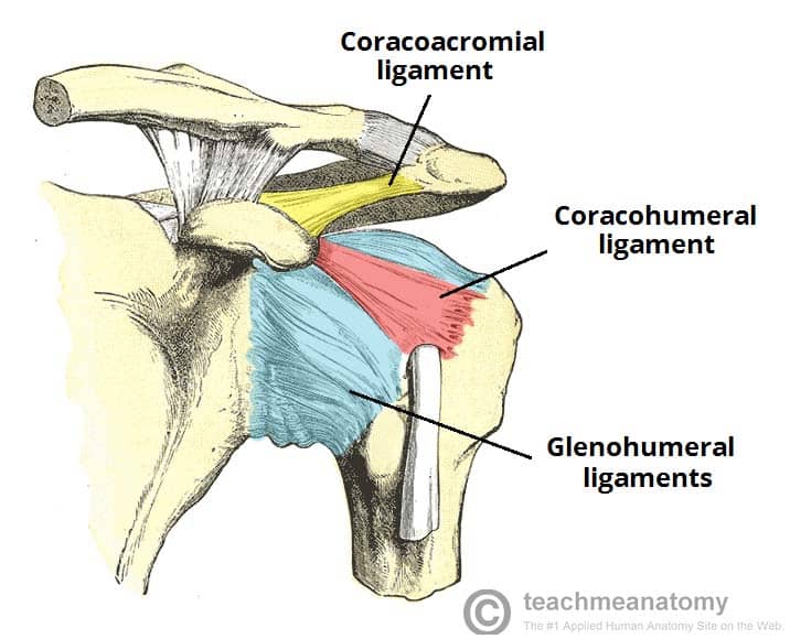
The Shoulder Joint Structure Movement Teachmeanatomy from teachmeanatomy.info The shoulder joint is formed where the humerus (upper arm bone) fits into the scapula (shoulder blade), like a ball and socket. Injuries to the glenohumeral ligaments can occur with shoulder dislocation. Diagram of the human shoulder joint. 8 name the arteries and the. Ebraheim's educational animated video describes muscle anatomy of the shoulder girdle and anatomy of the shoulder joint.anatomy of the shoulder muscles a. Stretching or tearing them can make your joints unstable. Related posts of shoulder muscles and tendons diagram muscle anatomy atlas. Learn vocabulary, terms, and more with flashcards, games, and other study tools.
It contributes to shoulder stability and, when.
Diagram of the human shoulder joint. Your upper arm bone (humerus), your shoulder blade (scapula), and your collarbone (clavicle). 8 name the arteries and the. The most flexible joint in the entire human body, our shoulder joint is formed by the union of the humerus, the scapula (or shoulder blade), and the clavicle (or collarbone). Once the ligaments, tendons, and muscles around the shoulder become loose or torn, dislocations can occur repeatedly. The shoulder joint is composed of the glenoid (the shallow shoulder socket) and the head of the upper arm bone known as the humerus (the ball). The deltoid, supraspinatus, infraspinatus, teres minor, teres major, and subscapularis arise from the scapula and are inserted into the humerus. Shoulder ligament tendon high resolution stock photography and images alamy : Related online courses on physioplus. Stretching or tearing them can make your joints unstable. Other important bones in the shoulder include: 3d video anatomy tutorial on the shoulder joint. Diagram of shoulder muscles and tendons.
Shoulder muscles and chest human anatomy diagram pdf arm vertebrate anatomy britannica shoulder anatomy diagram. Diagram of shoulder muscles and tendons.